Dentistry Illustrated
Long before the modern age of dentistry, oral issues presented a terrifying scenario. They [...]

Long before the modern age of dentistry, oral issues presented a terrifying scenario. They [...]
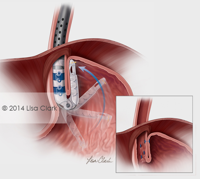
Esophagogastric fundoplication procedure to relieve chronic acid reflux caused by gastroesophogeal valve defect. The post Esophagogastric Fundoplication appeared first on Clark Medical Illustration.
James Archer is a medical illustrator and animator who combines artistic imagination with [...]
This is a medical-legal piece showing a dissection of the internal carotid artery. In this instance the word dissection does not refer to cutting something up to study. Here it is a tearing away of the interior wall of the carotid artery due to a spontaneous or traumatic injury. While once considered uncommon, spontaneous dissection is now known […]
One of my newest works is an eye cross section from above (superior) and from behind (posterior) that I finished a couple weeks ago. I also included the planning sketch. The thickness of each layer is very important to the overall accuracy and a lot of time was spent in the sketching stage to make […]
Pediatricians manage the physical, behavioral, and mental health of children from birth. They are [...]
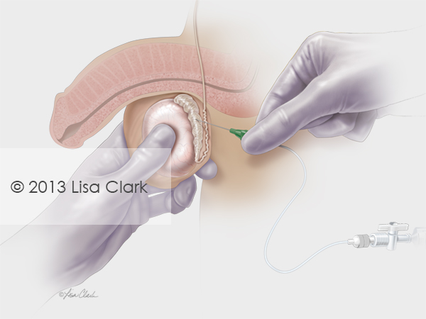
Testicular sperm aspiration allows physicians to aspirate epididymal fluid directly from the patient for male fertility testing or storage. The post Testicular Sperm Aspiration (TESA) appeared first on Clark Medical Illustration.
I’m often asked whether I draw by hand or use Photoshop. The answer is simply that I draw – sometimes on the computer and sometimes on paper. Photoshop is just another tool to put marks on paper, or in this case, pixels on the screen. I use a […]
The post Did You “Photoshop” That? appeared first on Clark Medical Illustration.
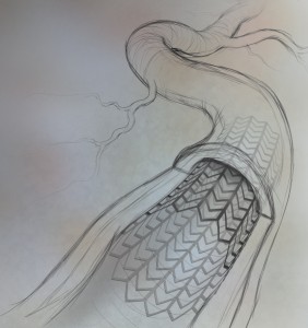
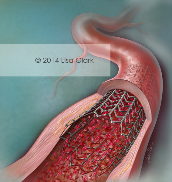
Stents are used to treat coronary artery disease and allow blood to flow through the artery. This image was created for a cardiology journal. The post Coronary Artery Stent appeared first on Clark Medical Illustration.
2014 Vesalius Trust Foundation Live Auction The Fairmanators Mission Brief http://www.crowdrise.com/thefairmanators What: Competitive Treasure Hunt (Live! At the 2014 AMI Meeting) When: Saturday, July 26th@ 6:30pm – 8:15pm Where: Downtown Rochester, MN – The Plaza Dear Fellow Fairmanator: This year, the VT Live Auction will consist of a competitive treasure hunt! Teams will unlock clues […]
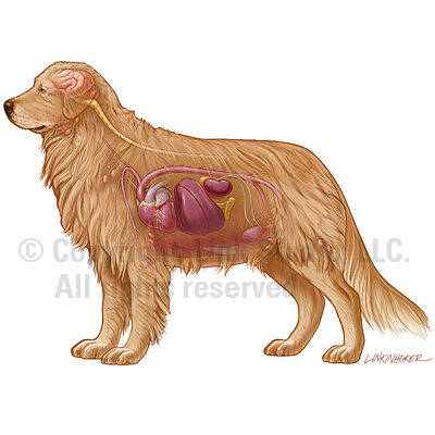
When the only means of communication come from painful whines or playful tail [...]
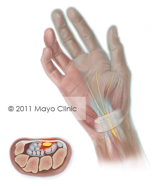
Carpal tunnel syndrome is a condition that causes numbness, tingling and burning in the hand. Carpal tunnel syndrome is caused by inflammation around the nerves in the wrist. The post Carpal Tunnel Syndrome appeared first on Clark Medical Illustration.
This editorial illustration was created for a July 2014 cover of American Family Physician. The image summarizes Constipation in Children. Childhood constipation is common and almost always functional without an organic etiology. Stool retention can lead to fecal incontinence in some patients. Often, a medical history and physical examination are sufficient to diagnose functional constipation. […]
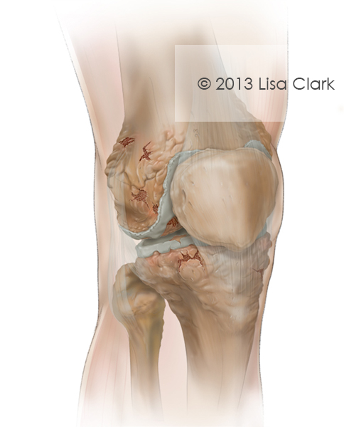
Arthritis is an inflammation of joints. Osteoarthritis (OA) is divided into stages. The highest stage, 4, is assigned to severe OA. OA that has become this advanced is likely causing significant pain and disruption to joint movement.
The post Severe Osteoarthritis of the Knee appeared first on Clark Medical Illustration.
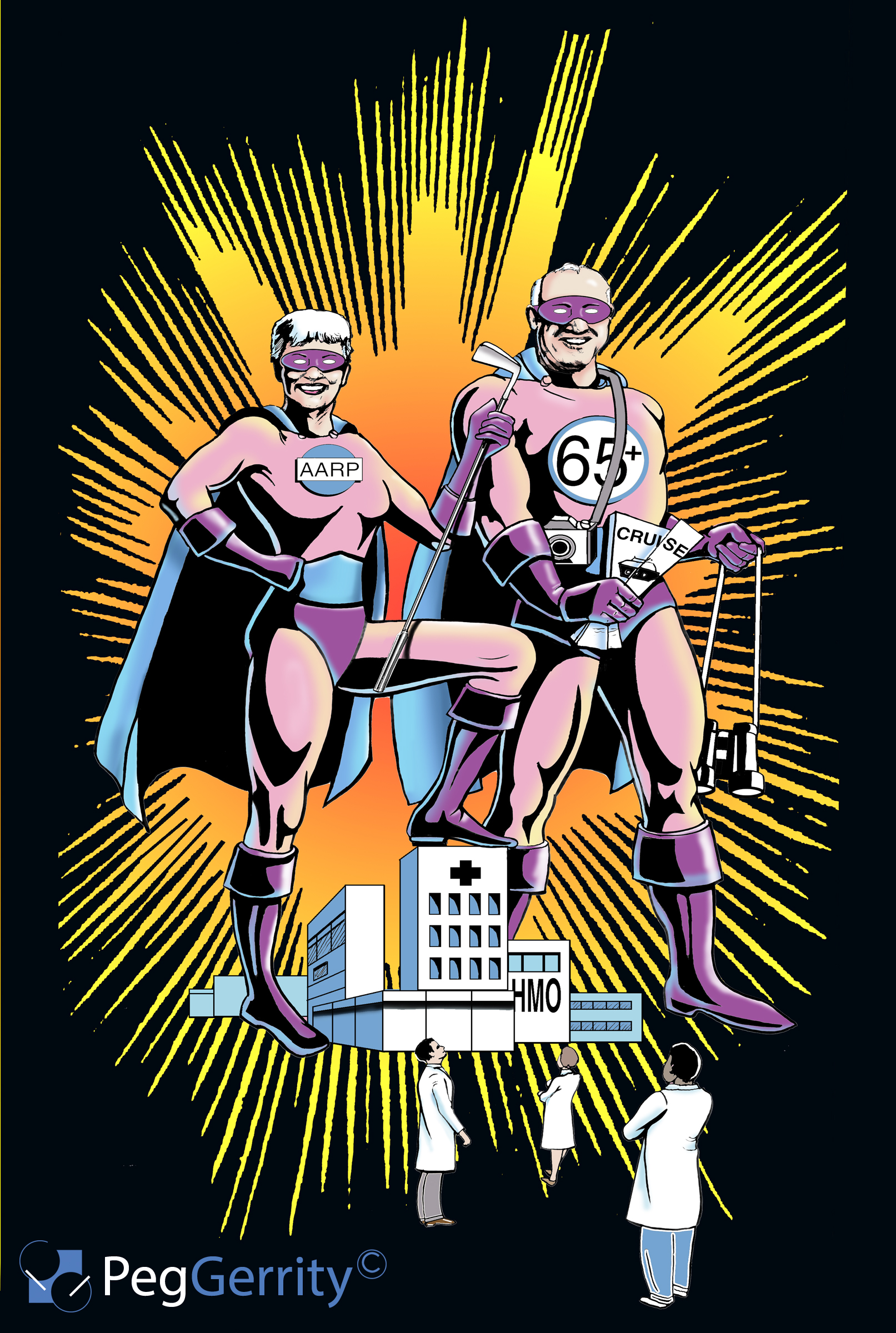
Today's aging population has had a profound effect on the healthcare industry, as [...]
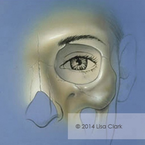
The illustration demonstrates desired position and proportion of the eye, brow and eyelid relative to the ocular orbit. The post Periocular Aesthetics appeared first on Clark Medical Illustration.
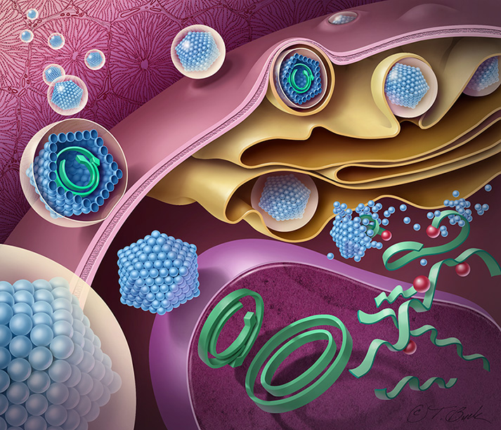
Todd is president and owner of Todd Buck Illustration, Inc. Todd Buck [...]
The pelvic floor is made of several muscle layers that form a bowl shape within the pelvis. This image illustrates a side view of the uterus, bladder, rectum and pelvic floor muscles. The post Female Pelvic Floor appeared first on Clark Medical Illust...
This is a still from a recent animation project on the feline olfactory system and appetite. The post Feline Olfactory System appeared first on Clark Medical Illustration.
Carotid stenosis is a condition in which the carotid arteries become narrowed or blocked. This image was used for a medical journal cover.
The post Carotid Stenosis appeared first on Clark Medical Illustration.
The hiatus is an opening in the diaphragm (the muscular wall separating the chest cavity from the abdomen). Typically, the esophagus goes through the hiatus and attaches to the stomach. In a hiatal hernia the stomach bulges up into the chest through that opening. This illustration depicts a hernia […]
The post Hiatal Hernia Graft appeared first on Clark Medical Illustration.
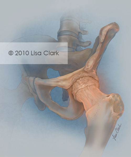
Osteoarthritis occurs when inflammation and injury to a joint cause cartilage tissue to break down. The breakdown of cartilage causes pain, inflammation, and deformity. The post Osteoarthritis of the Hip appeared first on Clark Medical Illustration.
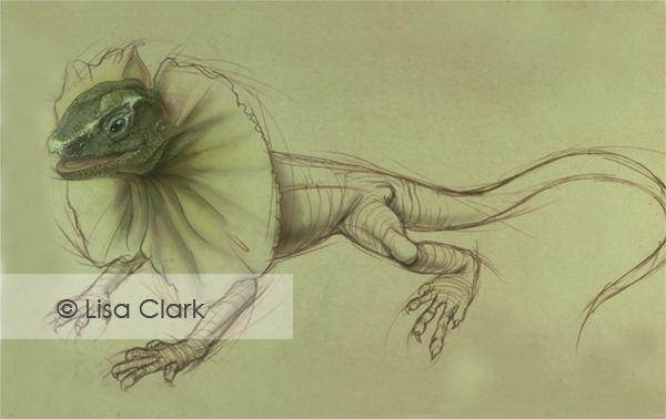
Chlamydosaurus have a frill of folded skin that normally lies around the back of the head that is erected when the lizard is threatened. These bipedal lizards can run very quickly and sometimes appear to be walking on water.
The post Frill Neck Lizard appeared first on Clark Medical Illustration.
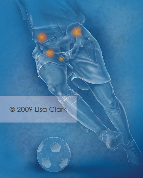
Apophyseal avulsion fractures of the hip and pelvis are injuries that usually occur in the adolescent athlete. This illustration was used for a medical journal cover.
The post Apophyseal Avulsion Fractures of the Hip and Pelvis appeared first on Clark Medical Illustration.
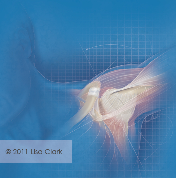
This illustration was used as the journal cover art for an article on healthy athletes with a history of shoulder instability.
The post Shoulder Instability in Young Athletes appeared first on Clark Medical Illustration.
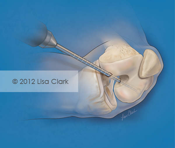
A femoral tunnel was created to aid with anterior cruciate ligament (ACL) reconstruction. This image was created for an orthopedic journal cover. The post Femoral Tunnel Drilling for ACL Reconstruction appeared first on Clark Medical Illustration.
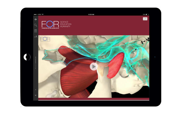
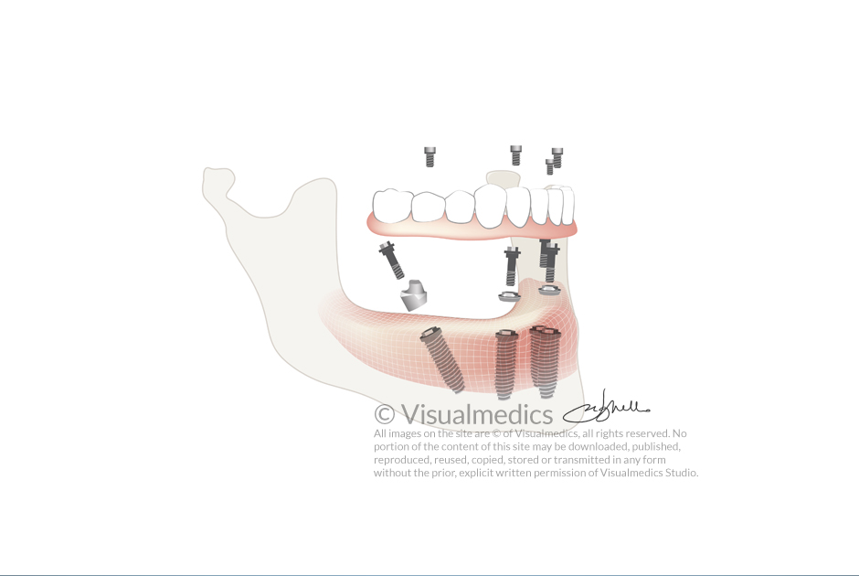
We had the pleasure to work with FOR and bring to life an exceptional resource for those desiring in-depth knowledge of temporomandibular joint anatomy. An incredible collection of images further enhances the experience of understanding the more intricate anatomic characteristics of the TMJ. The Temporomandibular Joint joins the existing digital textbook Single Implants and Their Restoration.
Wow can medical infographics help with health explanations? From prehistoric times to the digital age infographics have helped in comprehending various subjects. Medical infographics contribute greatly to today’s medicine and science. The first maps were created thousands of years before writing. Maps have been found drawn on the walls of Turkish city Çatal Hüyük in 6200BC. Christopher Scheiner used graphics to illustrate his astronomy research about the Sun and in 1786 William Playfair published his first book with statistical
I was recently given the opportunity to work in an unusual media — a large scale chalk mural at Lark on the Park restaurant in Dallas. Lark has a great interior design concept, plus some seriously amazing food. The restaurant walls are covered with blackboards and each […]
The post Making Murals appeared first on Clark Medical Illustration.
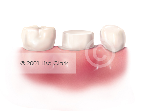
Crown: Tooth Prepared for Crown The post Crown: Tooth Prepared for Crown appeared first on Clark Medical Illustration.
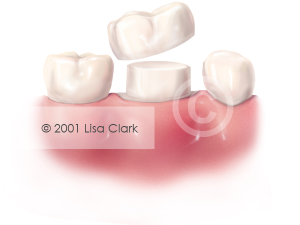
Crown: White Crown Near Final Position The post Crown: White Crown Near Final Position appeared first on Clark Medical Illustration.
Crown: White Crown in Final Position The post Crown: White Crown in Final Position appeared first on Clark Medical Illustration.
Crown: Gold Crown in Final Position The post Crown: Gold Crown in Final Position appeared first on Clark Medical Illustration.
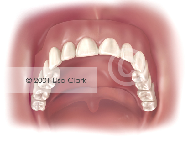
Dentures: Full Dentures in Place The post Dentures: Full Dentures in Place appeared first on Clark Medical Illustration.
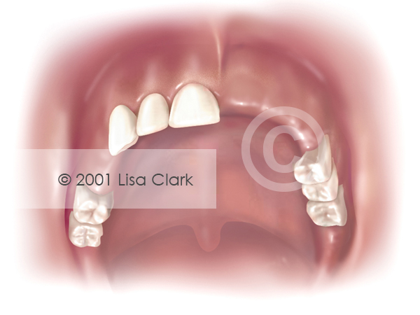
Dentures: Missing Teeth The post Dentures: Missing Teeth appeared first on Clark Medical Illustration.
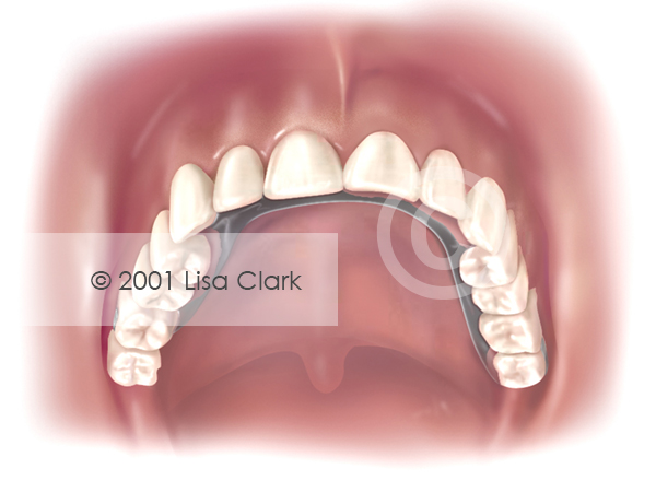
Dentures: Partial Dentures The post Dentures: Partial Dentures appeared first on Clark Medical Illustration.
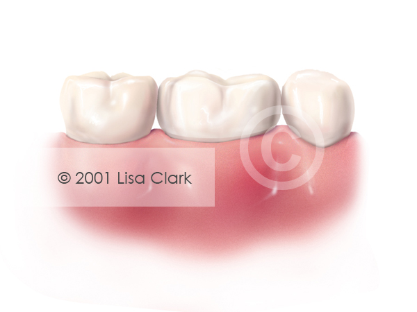
Teeth on either side of missing tooth have drifted toward the space, lower tooth has drifted upward into space. The post Drifting appeared first on Clark Medical Illustration.
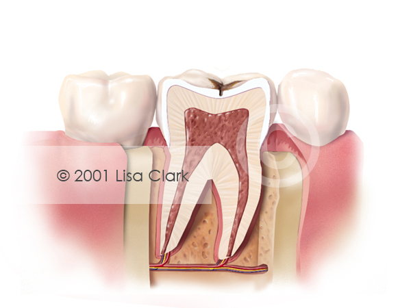
Progressive tooth decay. The post Dental Fillings 1: Progressive Tooth Decay appeared first on Clark Medical Illustration.
Tooth prepared for filling. The post Dental Fillings 1: Tooth Prepared for Filling appeared first on Clark Medical Illustration.
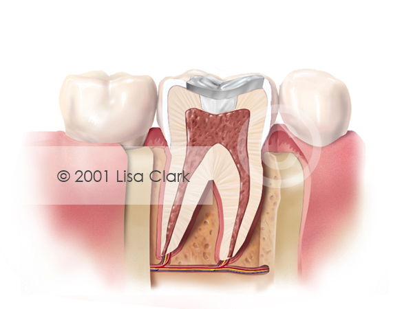
Dental Fillings 2: Tooth Filled with Silver Filling Material The post Dental Fillings 2: Tooth Filled with Silver Filling Material appeared first on Clark Medical Illustration.
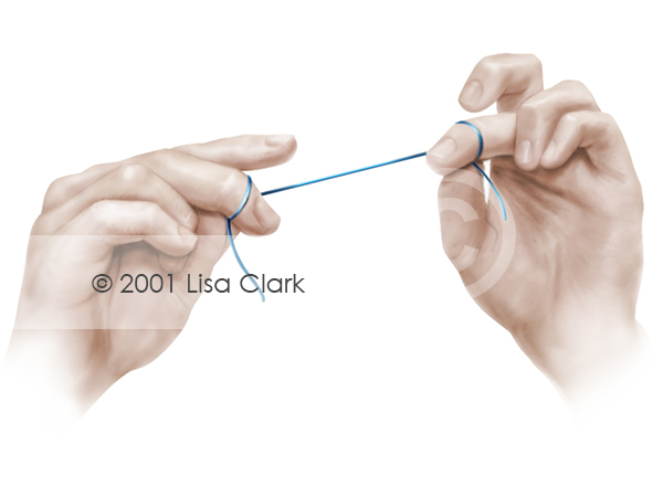
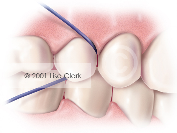
Dental Home Care: Holding Floss The post Dental Home Care: Holding Floss appeared first on Clark Medical Illustration.
Dental Home Care: Floss Properly Placed Between Teeth The post Dental Home Care: Floss Properly Placed Between Teeth appeared first on Clark Medical Illustration.
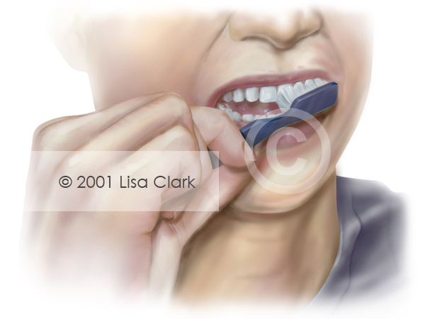
Dental Home Care: Proper Brush Placement The post Dental Home Care: Proper Brush Placement appeared first on Clark Medical Illustration.
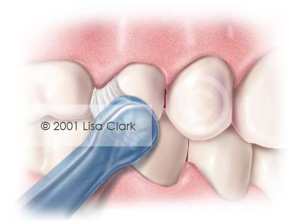
Dental Home Care: End Tuft Brush Properly Placed Next to Teeth The post Dental Home Care: End Tuft Brush Properly Placed Next to Teeth appeared first on Clark Medical Illustration.
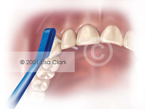
Dental Home Care: Proxi-brush Properly Placed Between Teeth The post Dental Home Care: Proxi-brush Properly Placed Between Teeth appeared first on Clark Medical Illustration.
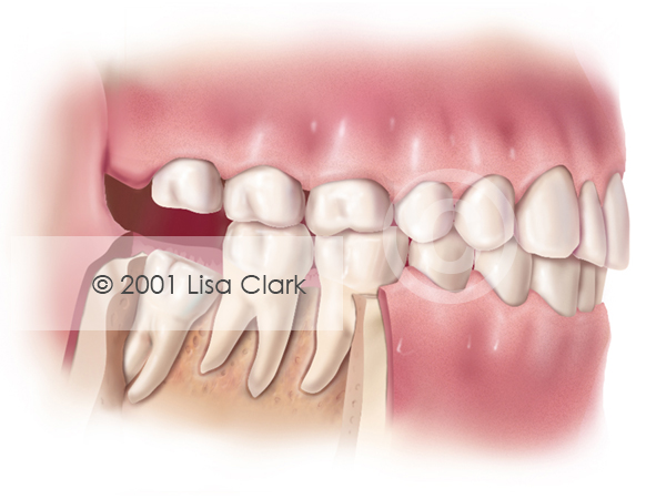
Impacted Wisdom Tooth: Soft Tissue Impacted The post Impacted Wisdom Tooth: Soft Tissue Impacted appeared first on Clark Medical Illustration.
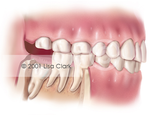
Impacted Wisdom Tooth: “Partial Bony” Impacted
The post Impacted Wisdom Tooth: “Partial Bony” Impacted appeared first on Clark Medical Illustration.
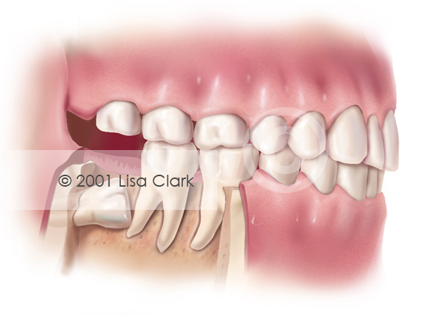
Impacted Wisdom Tooth “Full Bony” or “Horizontally” Impacted
The post Impacted Wisdom Tooth “Full Bony” or “Horizontally” Impacted appeared first on Clark Medical Illustration.
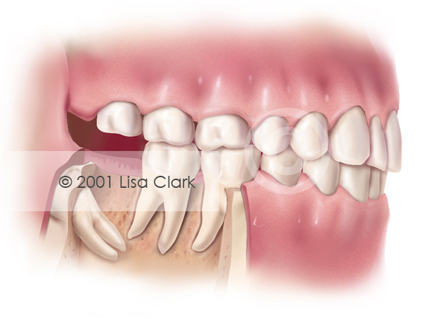
Impacted Wisdom Tooth: “Distally” Impacted
The post Impacted Wisdom Tooth: “Distally” Impacted appeared first on Clark Medical Illustration.
Absorb and metabolize these fantastic gastroenterology illustrations from the best medical artists in [...]
Dental Inlay: Tooth prepared for Inlay The post Dental Inlay: Tooth prepared for Inlay appeared first on Clark Medical Illustration.
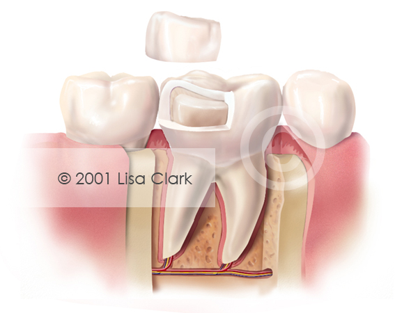
Dental Inlay: Inlay Near Final Position The post Dental Inlay: Inlay Near Final Position appeared first on Clark Medical Illustration.
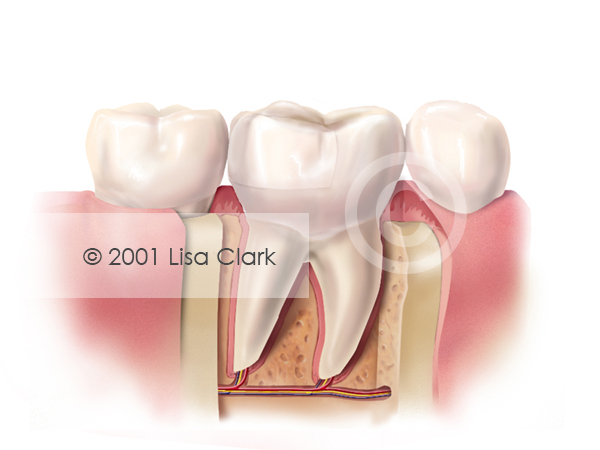
Dental Inlay: Inlay in Final Position The post Dental Inlay: Inlay in Final Position appeared first on Clark Medical Illustration.
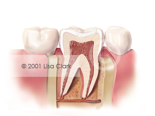
Dental Onlay: Onlay in Final Position The post Dental Onlay: Onlay in Final Position appeared first on Clark Medical Illustration.
Dental Onlay: Onlay Near Final Position The post Dental Onlay: Onlay Near Final Position appeared first on Clark Medical Illustration.
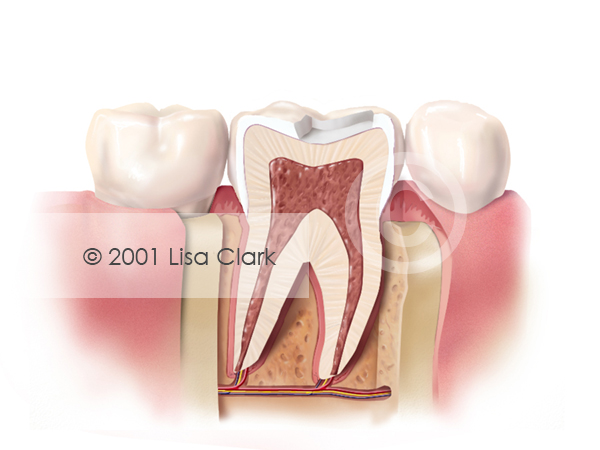
Dental Onlay: Tooth Prepared for Onlay The post Dental Onlay: Tooth Prepared for Onlay appeared first on Clark Medical Illustration.
Dental Post 1: Build Up Material in Place
The post Dental Post 1: Build Up Material in Place appeared first on Clark Medical Illustration.
Dental Post 1: Prefabricated Post in Place
The post Dental Post 1: Prefabricated Post in Place appeared first on Clark Medical Illustration.
Dental Post 1: Damaged Tooth The post Dental Post 1: Damaged Tooth appeared first on Clark Medical Illustration.
Dental Retention Pin The post Dental Retention Pin appeared first on Clark Medical Illustration.
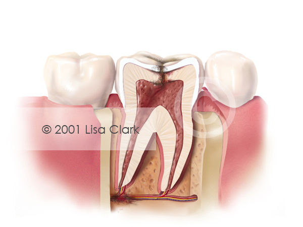
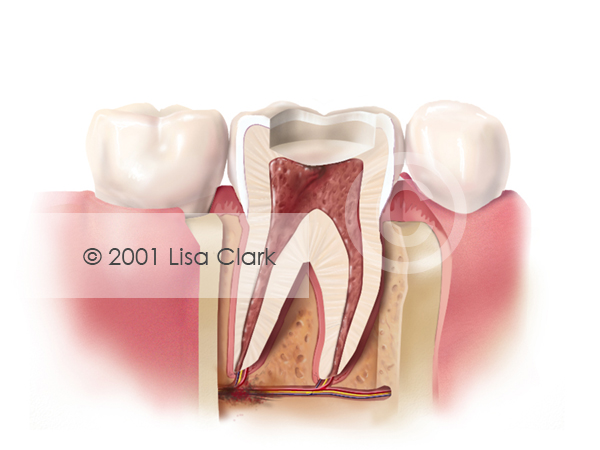
Root Canal: Tooth Requiring Root Canal The post Root Canal: Tooth Requiring Root Canal appeared first on Clark Medical Illustration.
Root Canal: Enamel and Dentin Removed The post Root Canal: Cross Section of Tooth Requiring Root Canal appeared first on Clark Medical Illustration.
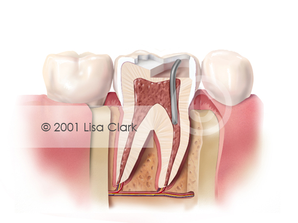
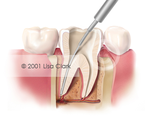
Root Canal: Pulp Removed The post Root Canal: Pulp Removed appeared first on Clark Medical Illustration.
Root Canal: Canals Filled The post Root Canal: Canals Filled appeared first on Clark Medical Illustration.
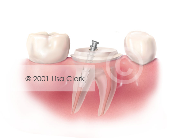
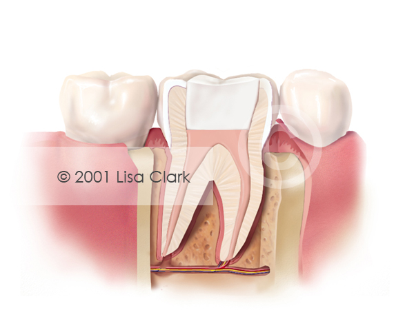
Root Canal: Final Filling (White Material) The post Root Canal: Final Filling (White Material) appeared first on Clark Medical Illustration.
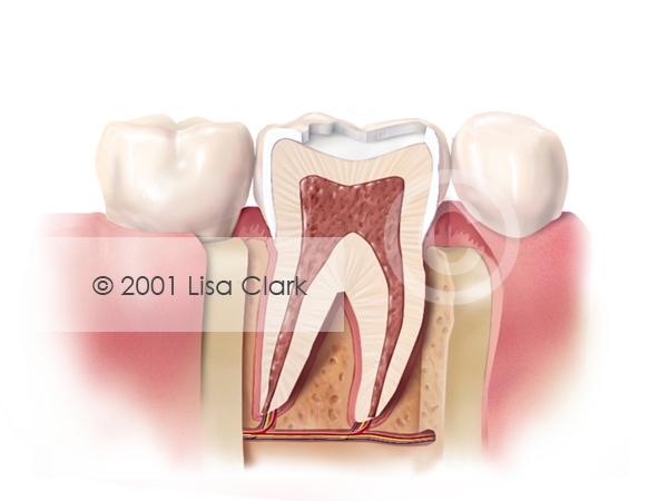
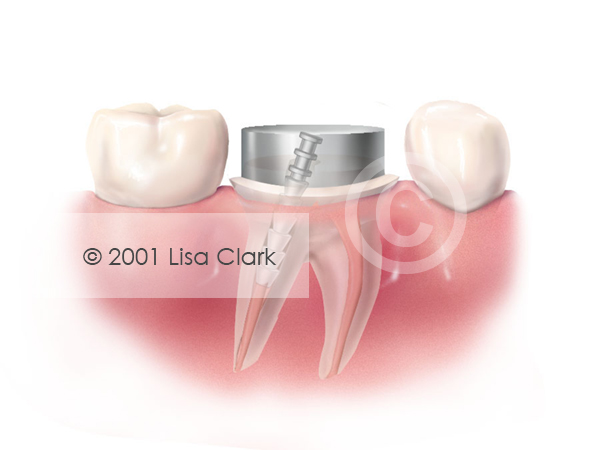
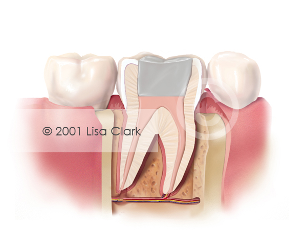
Root Canal: Final Filling (Silver Material) The post Root Canal: Final Filling (Silver Material) appeared first on Clark Medical Illustration.
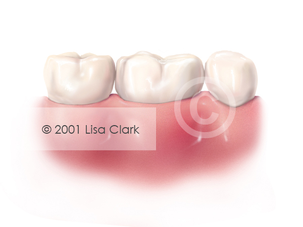
Sealants: Finished Procedure The post Sealants: Finished Procedure appeared first on Clark Medical Illustration.
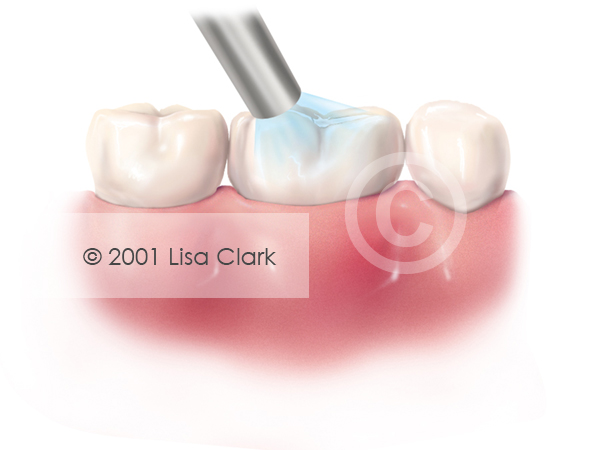
Sealants: Sealant Material Cured by Light The post Sealants: Sealant Material Cured by Light appeared first on Clark Medical Illustration.
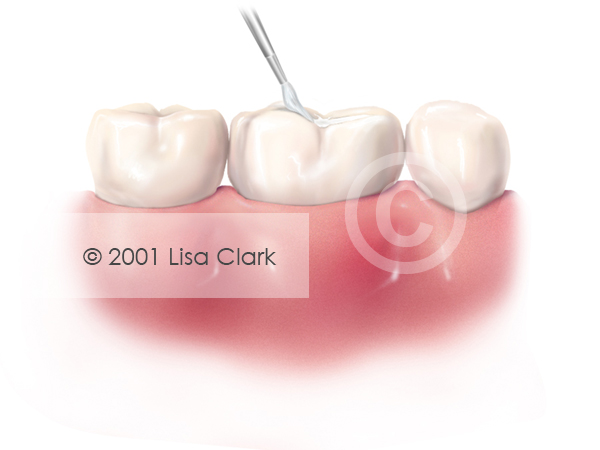
Sealants: Sealant Material Applied to Grooves The post Sealants: Sealant Material Applied to Grooves appeared first on Clark Medical Illustration.
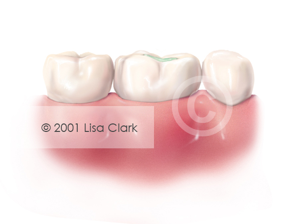
Sealants: Etchant Applied The post Sealants: Etchant Applied appeared first on Clark Medical Illustration.
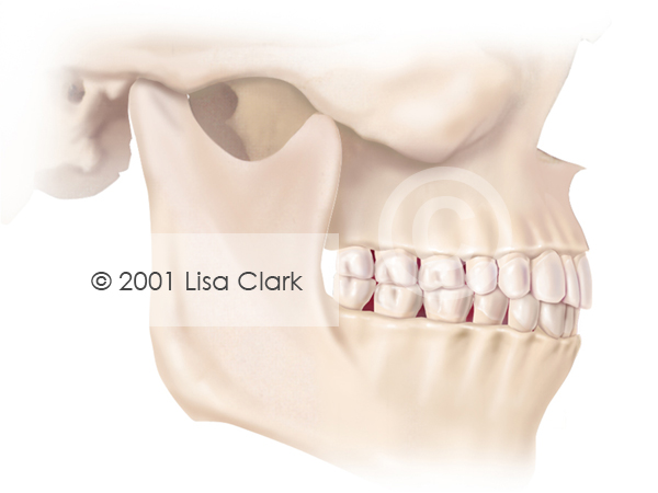
TMJ Dysfunction: Normal Temporamandibular Joint The post TMJ Dysfunction: Normal Temporamandibular Joint appeared first on Clark Medical Illustration.
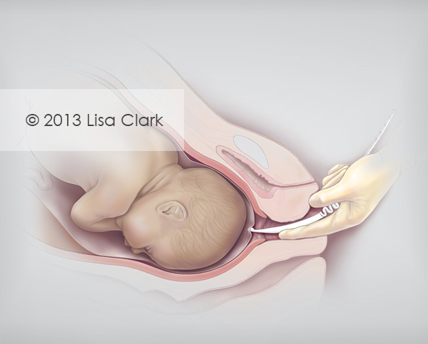
Amnion perforators are used to puncture the amniotic sac (amniotomy) during labor. This illustration was used for a medical device manufacturer’s marketing materials.
The post Amniotic Membrane Perforator appeared first on Clark Medical Illustration.
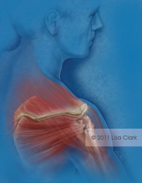
A rotator cuff tear is a tear in one of the tendons of the four rotator cuff muscles in the shoulder. This illustration was used as a journal cover illustration demonstrating the repair of a u-shaped rotator cuff tear. The post Rotator Cuff Tear appea...
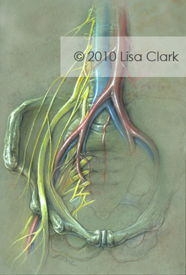
Illustration of lumbo-sacral nerve plexus, pelvis, iliac arteries, femoral arteries and lumbar spine. The post Sacral Nerve Plexus appeared first on Clark Medical Illustration.
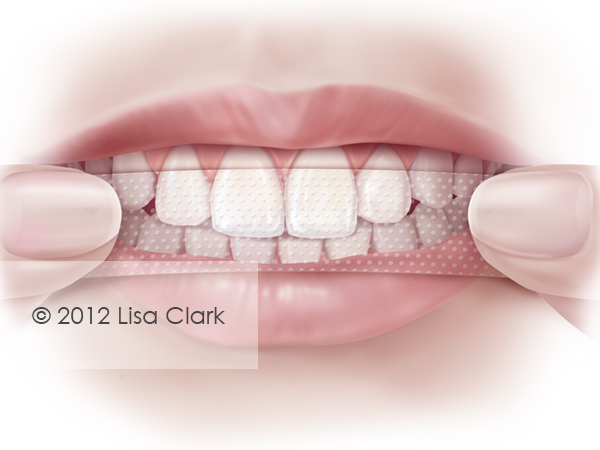
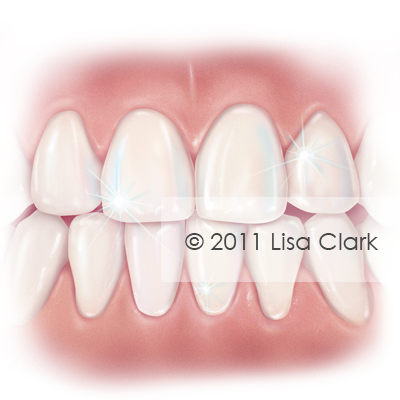
Teeth whitening strips are a popular home remedy for fixing stained or discolored teeth. This image was created as a marketing piece for a dental product manufacturer.
The post Teeth Whitening Strips appeared first on Clark Medical Illustration.
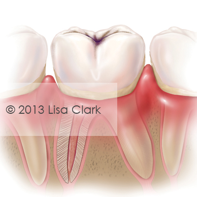
Buccal (frontal) view of molar with dental decay. The post Dental Caries (Cavities) appeared first on Clark Medical Illustration.
Percutaneous coronary intervention (PCI) for chronic total occlusions. The post Percutaneous Coronary Intervention appeared first on Clark Medical Illustration.
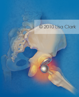
Dislocation is an uncommon, but potential complication to hip replacement surgery. This illustration demonstrates the failure of a metal on metal hip implant. The post Hip Replacement Failure appeared first on Clark Medical Illustration.
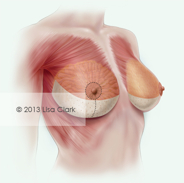
This image was used to demonstrate the placement of a new biological graft used in comprehensive breast reconstruction and revision procedures, including mastopexy or “breast lift”.
The post Mastopexy with Biological Graft appeared first on Clark Medical Illustration.
Frontal view of health adult teeth and gums. The post Healthy Teeth appeared first on Clark Medical Illustration.
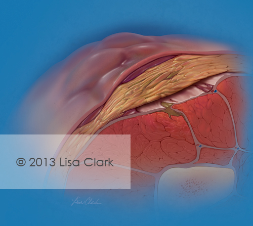
Necrotizing fasciitis (NF), commonly known as flesh-eating disease or Flesh-eating bacteria syndrome, is a rare infection of the deeper layers of skin and subcutaneous tissues, easily spreading across the fascial plane within the subcutaneous tissue.
The post Necrotizing Fasciitis appeared first on Clark Medical Illustration.
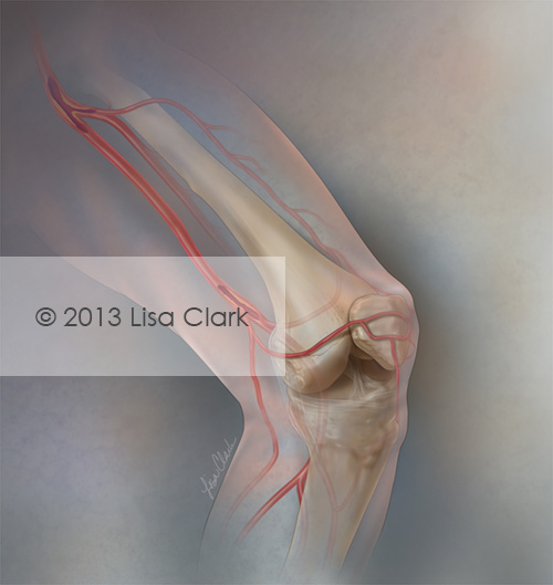
Journal cover illustration about the difficulties and options for the treatment of superficial femoral artery disease (SFA). The post Superficial Femoral Artery Disease appeared first on Clark Medical Illustration.
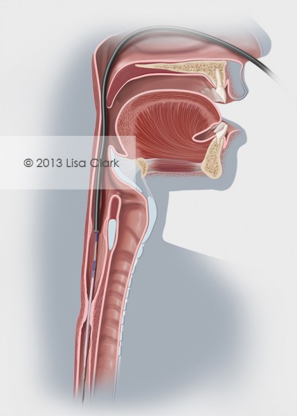
Esophageal stricture is a narrowing of the esophagus that causes swallowing difficulties. This image was used for medical device marketing materials.
The post Esophageal Stricture appeared first on Clark Medical Illustration.
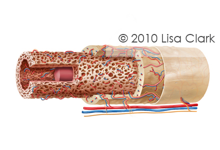
Long bone anatomy consists of a layer of connective tissue called the periosteum. The outer shell of the long bone is compact bone with a deeper layer of cancellous bone (spongy bone) which contains red bone marrow. The interior part of the long bone is the medullary cavity with the […]
The post Long Bone Anatomy appeared first on Clark Medical Illustration.
This editorial illustration was created for a May 2014 cover of American Family Physician. The image summarizes Unintentional Weight Loss in Elderly. Elderly patients with unintentional weight loss are at higher risk for infection, depression and death. The leading causes of involuntary weight loss are depression (especially in residents of long-term care facilities), cancer (lung […]
Diagnosis and Initial Management of Dysmenorrhea AMIMI S. OSAYANDE, MD, and SUARNA MEHULIC, MD, University of Texas Southwestern Medical Center, Dallas, Texas Am Fam Physician. 2014 Mar 1;89(5):341-346. Dysmenorrhea is one of the most common causes of pelvic pain. It negatively affects patients’ quality of life and sometimes results in activity restriction. A history and physical examination, including […]
Learn more about the new model in Cloud publishing, its practicality and editorial usage in medicine. Cloud publishing allows for faster information anywhere and on any device and is quickly becoming popular also for the medical and scientific publishing industry. A cloud publishing environment allows for remote access to programs, files and services on the internet, and it is possible to access these on any device with internet access. Google Docs is an excellent example
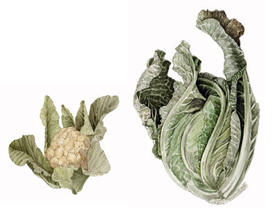
Fruits and vegetables with a little wine, conceptualized by the Medical Illustration Sourcebook [...]
Portable Organ Perfusion is a startup company innovating with a truly disposable portable organ perfusion system. We offered all support needed to build up the visual identity from scratch. The branding of the startup needed to be taken care thoroughly. So we set up appointments to understand the client’s needs and made up a strategy. We rolled into the team’s daily schedule to make it happen, time was not on the team’s side. Startups are constantly busy planning the latest
We recently concurred to start a exciting project. The Client looked for an illustration agency able to commit with sharp deadlines and listen to multiple associates feedbacks. We created the right recipe for a branded medical dental illustration collection supporting their content. The client is creating an online global reference portal illustrating procedures, making material available to professionals worldwide. The dental illustrations developed should aid the understanding of articles by providing a visual educational reference to the techniques used. We pitched with
The welcome return of spring also means the inevitable return of allergies. Pollen, [...]
Even though we're named the Medical Illustration Sourcebook, there are several more types [...]
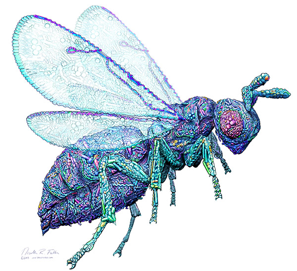
Explore the work of Nicolle Rager Fuller at medillsb.com! Renown for her natural [...]
Valentine's Day is the perfect opportunity to showcase the myriad of techniques and [...]
Our bodies are made up of the trillions of cells encoded by our unique DNA–right? Current research is diving into another rabbit hole, with suggestions that the microbes that coexist with us are just as much US, as our ‘own’ … Continue reading →