
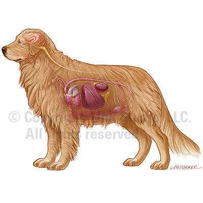
When the only means of communication come from painful whines or playful tail wags, accurate canine veterinary health illustrations are vital in understanding what is happening inside of our furry companions.
Check out the work of several Medical Illustration Sourcebook artists who are skilled not only in human anatomy, but also in a wide range of veterinary realms.


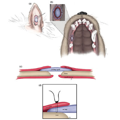
Illustrated steps of a free auricular cartilage graft for repair of an oronasal fistula in a canine. (a) Harvesting of auricular cartilage from caudal aspect of right ear pinna; (b) Ventral view of canine maxilla showing locations of ONF and placement of cartilage; (c) Cross-section view of cartilage in compartment between oral mucosa and hard palate; (d) Diagram of suture placement through oral mucosa and cartilage.\
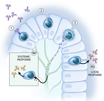
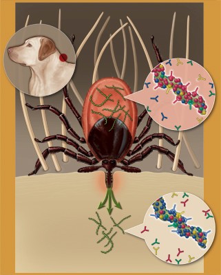
Illustration of the local and systemic immune response triggered by immunoglobulins in the lumen of the dog gut. Cells of lumen wall and antibodies shown.


