Featured Image: Alexander & Turner, Inc. 

Embryology has become an important research area for studying the genetic control of the development process, its link to cell signalling, its importance for the study of certain diseases and mutations, and in links to stem cell research.

Below we’ve compiled a selection of illustrations exploring different aspects of embryology and the reproductive process. Click on the illustrator’s name to see more of their work.

Steve Oh And Myriam Kirkman-Oh
KO Studios
KOStudios_CellDivision

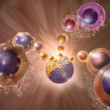
Stem Cells in the process forming a blastula


Laurie O’Keefe

DNA Illustrations, Inc.

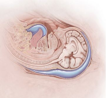
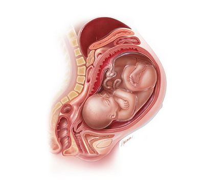
Midsagittal section of fetus in utero showing hydrops fetalis.


Linda Nye

Linda Nye

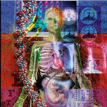
Large hanging panel incorporating elements of the past to future of medical visualization, for the UCSD Biomedical Library interior.


Joon Lee
CG TOKI

CGToki

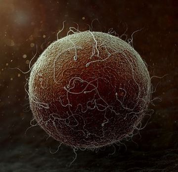
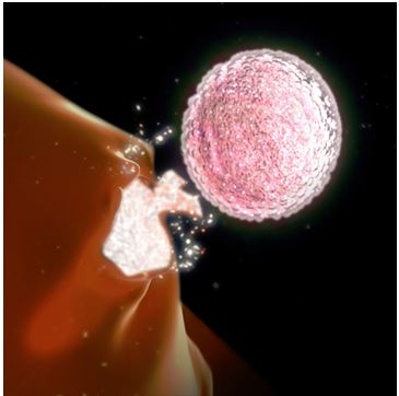
Human Egg Cell


Peg Gerrity

Peg Gerrity

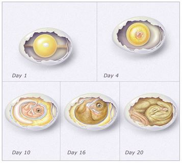
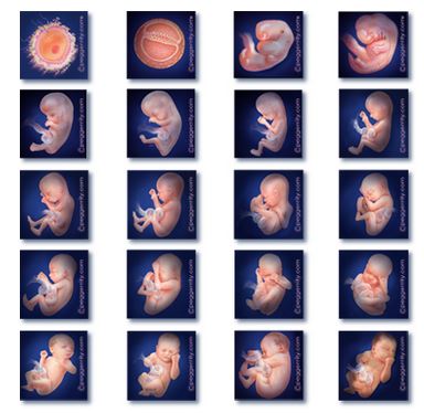
20 Biweekly images of human fetal development currently used by various US State Departments of Health to comply with the WRTK abortion legislation. Contact the illustrator if your department would like to lease these images. You may view larger samples at the Louisiana departmental website:http://new.dhh.louisiana.gov/index.cfm/page/986 (fetus foetus fetal foetal embryo embryology pregnancy pregnant abortion)
 

Jim Dowdalls

MartensKiefer

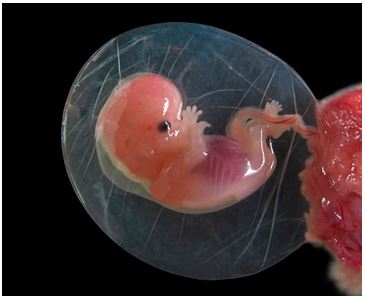
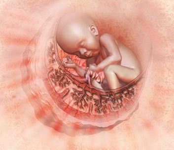
8 week fetus with amnion sac


Nicolle Rager Fuller
Sayo-Art LLC

Sayo-Art

A baby develops within a healthy placenta. Surpisingly, the placenta is not the mother’s organ, but grows from the cells first laid down by the fertilized egg. To successfully support the pregnancy, the placenta has to convince the mother’s immune system that it should be there. Scientists are studying this process to determine its involvement in various pregnancy complications
 

Matthew Chansky
Medical Design Agency

MatthewChansky

Illustration of uterine anomaly: septate uterus for medical legal demonstration


Phototake – Masters in Medical Images

Phototake

Female human embryo, seven weeks after fertilization. The fetus is enveloped in the amniotic sac and attached to the placenta. The umbilical cord size is 18mm


Beth Anderson
Arkitek Scientific

Ariketk

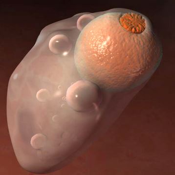
Still from a series of animations created for veterinary reproductive physiology courses. This image is of a cow ovary during the luteal phase

Edmond Alexander And Cynthia Turner
Alexander & Turner, Inc. 

Alexander & Turner

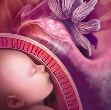
Womens’ reproductive health, with uterus, ovaries, ovulating ovary, and ovum traveling through the fallopian tube with its nourishing follicular cells


Delilah Cohn
The Medical Illustration Studio

Delilah Cohn

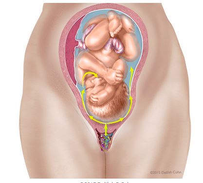
Illustration of a late term fetus in the womb showing how some bacteria in the birth canal can pass through the amniotic membrane into the fluid where it is inhaled by the fetus. Other drawings in the series show bacteria traveling through the fetal circulatory system to the brain.


Thomas A. Graves

ThomasAGraves


Bonnie Hofkin

Bonnie Hofkin


Polymime Studio
Polymime Animation Co.

Polymime Animation Co.

Egg


Lauren Hamm

Lauren Hamm

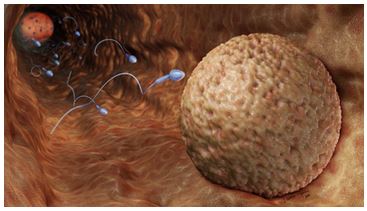
Fertilization