
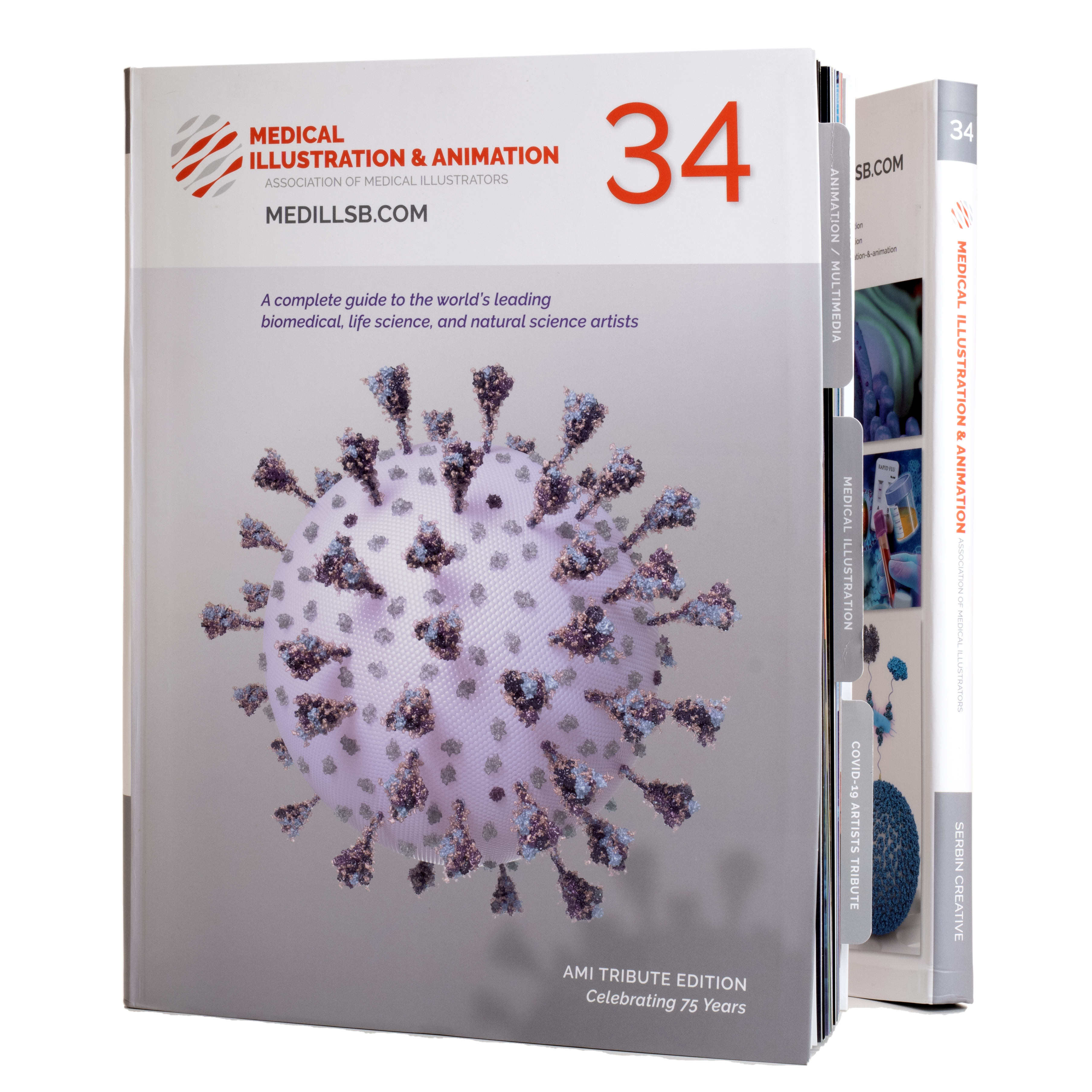
The AMI Tribute Edition: Celebrating 75 Years
We are proud to announce the release of the Medical Illustration & Animation Sourcebook No.34! As a complete guide to the world’s leading biomedical, life science and natural science artists, the Sourcebook is the number one resource for medical art buyers. Spanning over 170 pages, the Sourcebook introduces you to some of today’s most outstanding artists. They have the scientific background and expertise necessary to understand your project, media selection, and target audience. These incredible artists have a wealth of knowledge and a dedication to scholarship that stems from a lifetime of study.
75 Years of the AMI
This year’s edition is a very special tribute for the 75th anniversary of the Association of Medical Illustrators. The first gathering of the five founding members of the AMI was on July 10th, 1944. Seventy-five years later, the AMI is now comprised of over 800 members across 4 continents.
“Historically, our roots are in medical textbook and journal publishing. Now, as technology and digital media transform both science and communication, AMI members are on the leading edge of this dynamic multidisciplinary frontier.” – Association of Medical Illustrators
Click the button below to read the full history of the AMI.
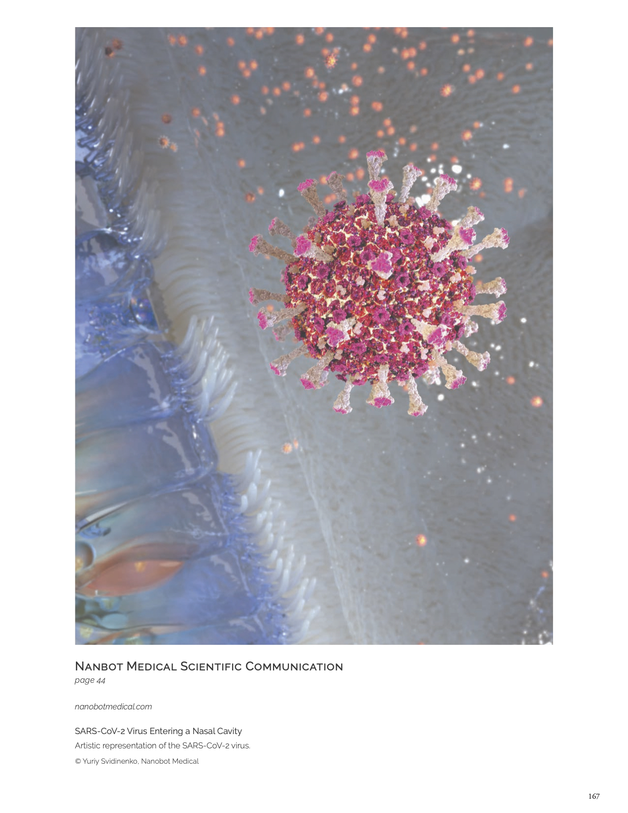
COVID-19 Tribute Section
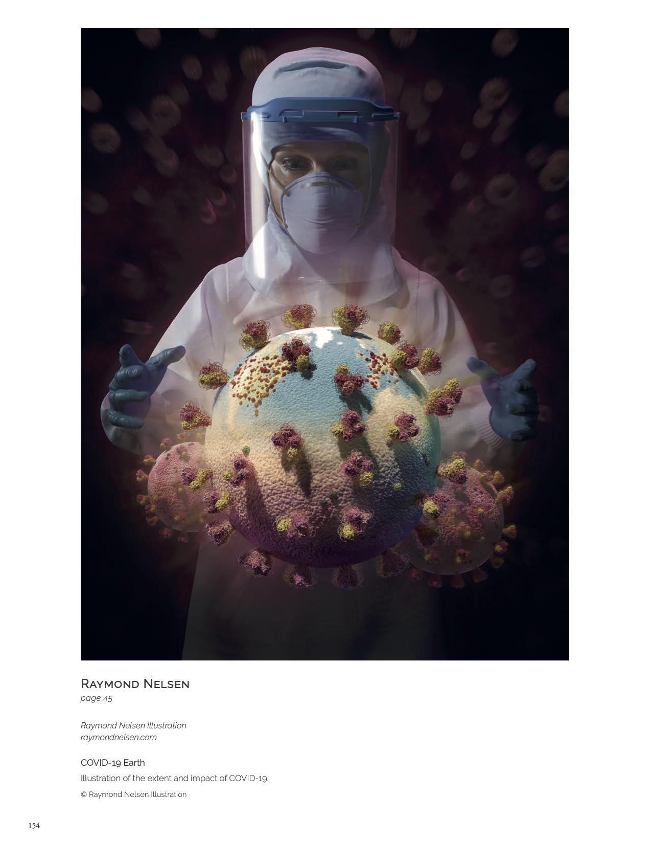
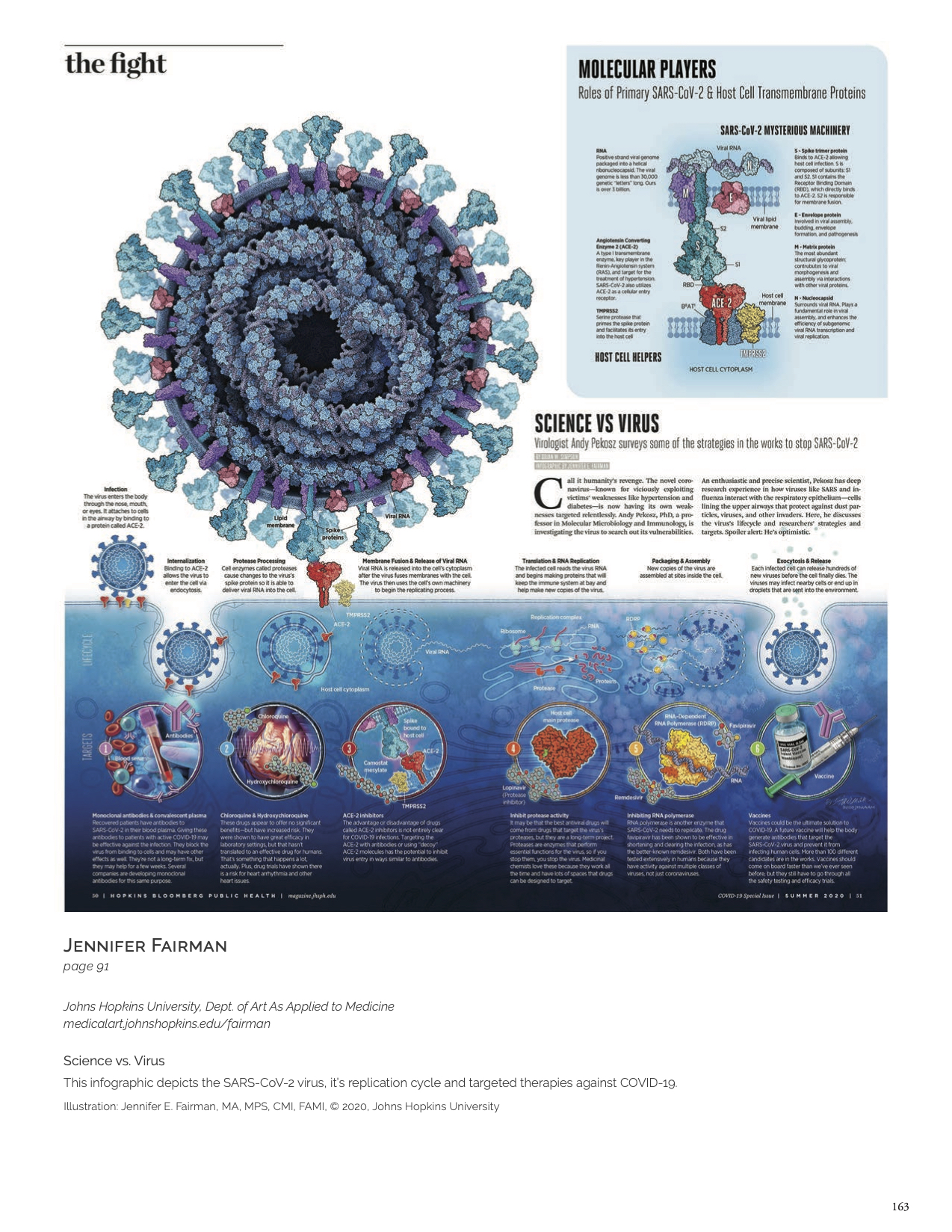
The production of this book occurred during the height of the pandemic, which made it clear the role that the artists in this book have had in making science accessible to everyone. Their efforts, energy, knowledge and expertise contributed immensely to the science of Covid. We decided to pay a special tribute to these amazing artists and their invaluable contributions.
“Medical illustrators raced to collaborate with researchers to help uncover the structure of the novel coronavirus COVID-19…This was a massive community undertaking to educate the public and attempt to save lives. Medical illustrators were proud to be part of this fight, hoping our contributions helped to stem the tide of mounting infection rates and public anxiety. We shared research, newly released 3D structural models of the envelope proteins, and everything else we discovered about the virus’s structure and mechanics in an open-source repository. The images we created have helped define our collective experience and visual memories of this historic time.”
– Rachel Bajema, MS, CMI, Editor
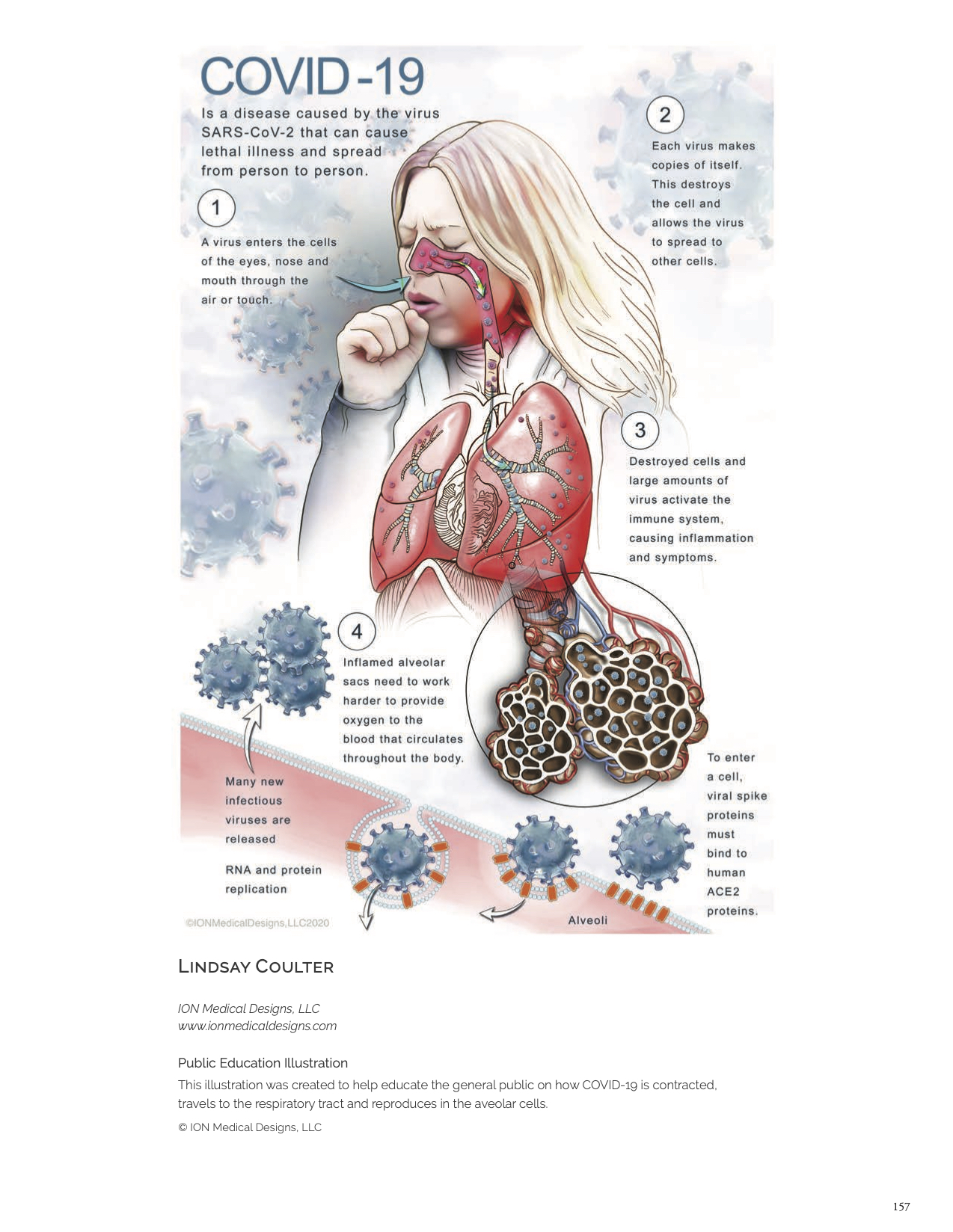
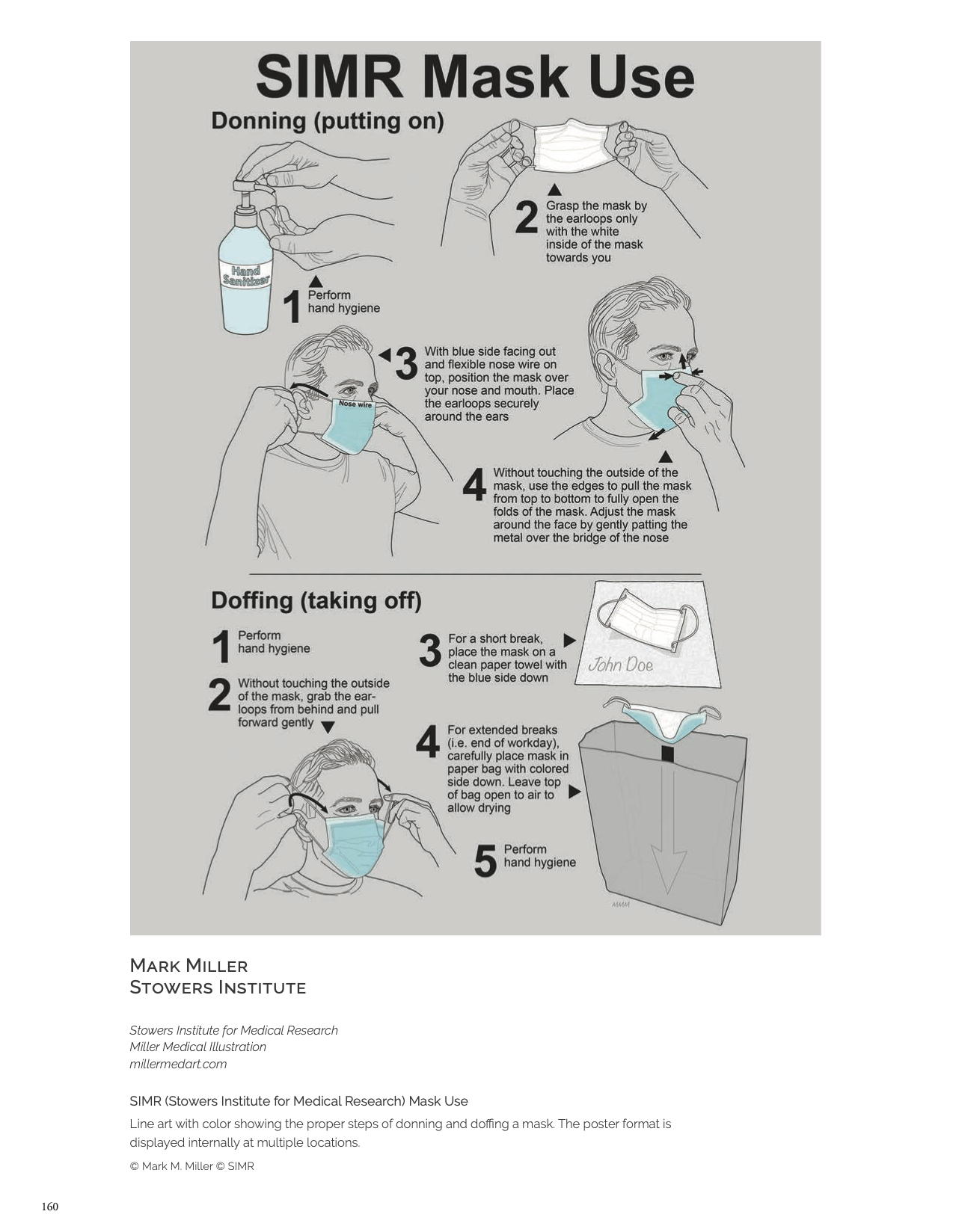
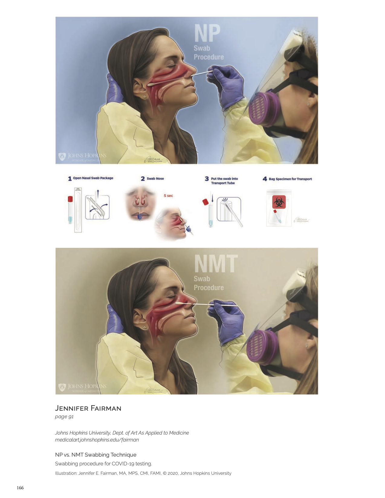
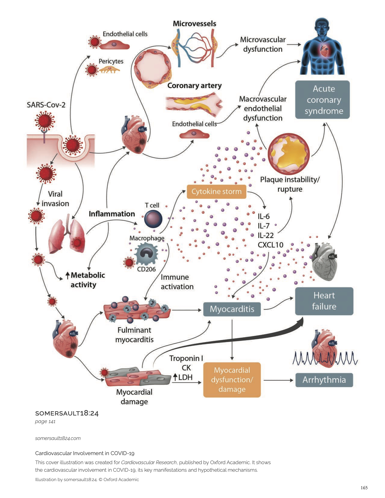
We put out a call for artists to share their Covid work and they surely delivered: Everything from awareness posters, scientific renderings of the research as it developed, infographics of the form and shape of the virus, how to wear a mask, how it affects the lungs, infographics of how it affects the body, how the vaccine works and instructional graphics for hospitals on how to administer nasal swabs. Their flood of submissions really brought to light that medical illustrators are more than illustrators – they help society and the betterment of humanity.
“We look to 2021 with hope, celebrating science that has provided protection and served humankind, and continuing to ask hard questions, search for new ideas, and explaining the best of these through art.” – Rachel Bajema, MS, CMI, Editor
View the digital book now to see their work! Entries start on page 152.
Cover Artist: Melanie Connolly
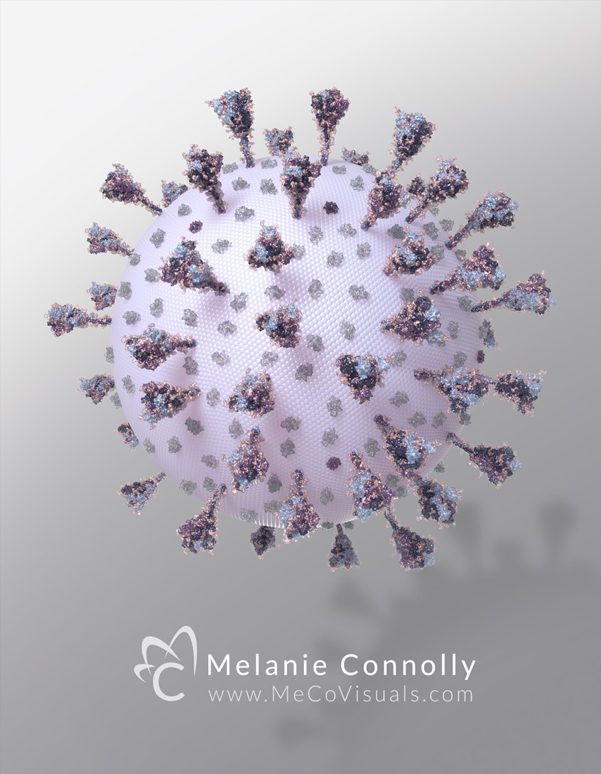
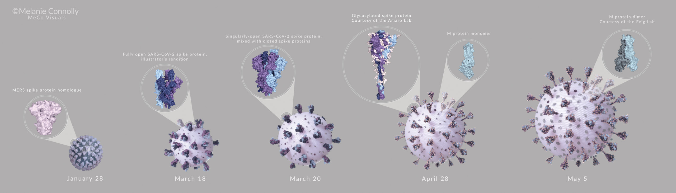
From the Artist: “What a difference collaboration makes! When SARS-CoV-2 began to spread worldwide, Medical Illustrators rushed to build accurate visuals with current information. While we regularly work on the leading scientific edge, this situation was different. As new research was published daily, medical artists were challenged to efficiently and quickly evaluate hundreds of sources. Michael Konomos started an editable document, and Medical Illustrators put their brains together. The resulting visuals changed over time, much like RNA viruses themselves.
I started this viral image with MERS spikes as opposed to SARS-CoV-2 spikes, which were added later once their structure had been elucidated and made available. As new updates to the protein databank and our understanding of the surface proteins evolved, various components, like the E and M proteins, were added to increase the accuracy of the image. Eventually, glycosylated spike proteins were added courtesy of the Amaro Lab, and dimerized M proteins were added courtesy of the Feig Lab.
This compilation is in honor of my medical illustration colleagues. Without their feedback, late phone chats, and Twitter messages the resulting image would have been very different— and not nearly as fun to make.
Special thanks: M. Konomos, V. Falconieri, D. Goodsell, J. Fairman, E. Maizels, R. Amaro, M. Maritan, Feig Lab, S. Bond, P. Gerrity, and all who contributed.” – Melanie Connolly
About the Artist: Melanie Connolly is a Medical Illustrator and 3D Medical Animator with The University of Texas Medical Branch at Galveston in the Department of Surgery. Through her companies MeCo Visuals and Chicago Medical Graphics, she also provides visual communication in the fields of MedTech, Pharma, and pediatric healthcare education. She recently served as the Marketing Director with the Austin Healthcare Council, focusing on local and international healthcare collaboration and transformation.


