
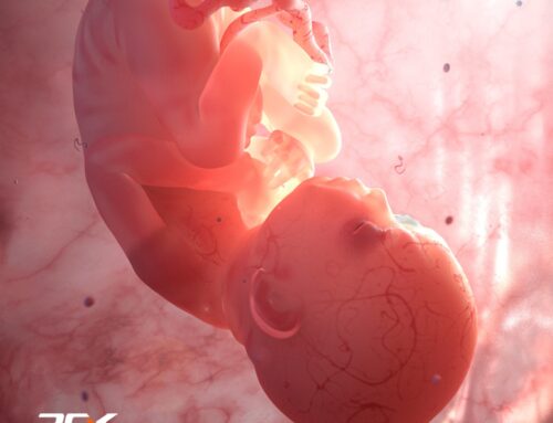
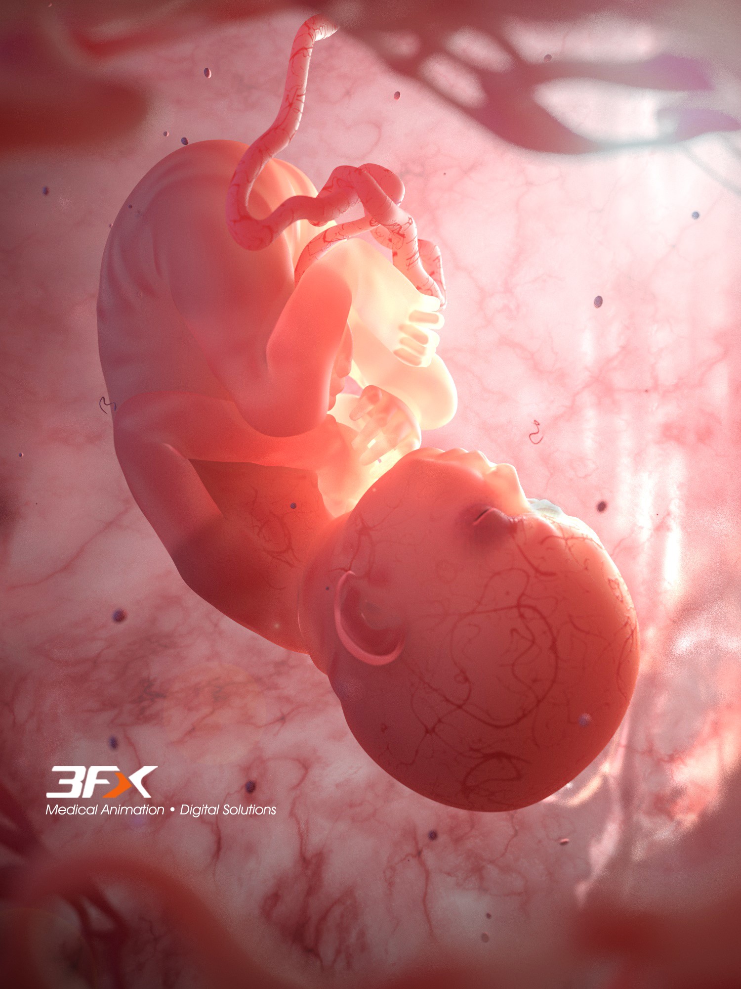
Featured Image ©Fran Milner
The eye is the organ of vision. It allows us to see and interpret the shapes, colors, and dimensions of objects in the world by processing the light it reflects or emits. The human eye is also an immensely complex organ with the possibility of errors and various types of injury, infection, and changes due to systemic disease.
Since the eye allows us to visually communicate with the world around us, studying its intricacies is a very important aspect of modern medicine.
Here are a few studies into visual communication, illustrated by the Medical Illustration Sourcebook Artists.
Audra Geras | Geras Healthcare Productions

The Healthy Eye
Gary Carlson
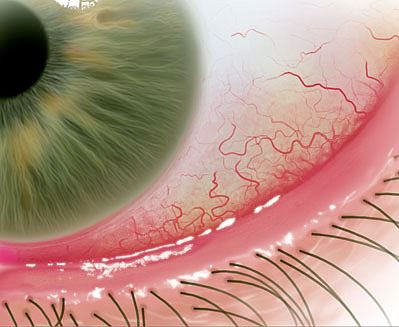
Dry eye leads to red and irritated sclera
Fran Milner
Sectioned view of the normal eye, detailing main structures
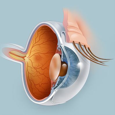
Ben Smith
Eye interior
Elaine Kurie
Tear film
Science Source
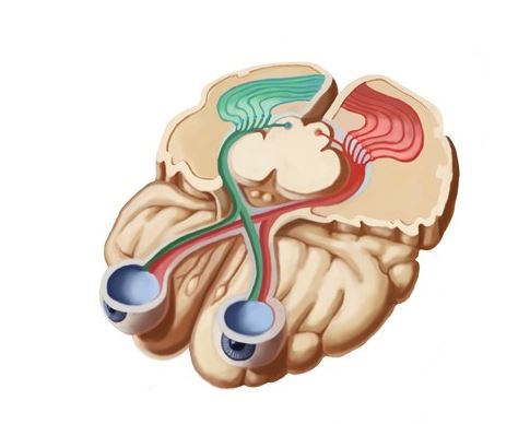
Visual pathways from the retina of the eye to the brain cortex
Joan Kozel
Nerves of the Eye
Richard Weaver | DWeaver Designs-BioMedical Illustration
Eye cross-section
Erica Beade | MBC Graphics
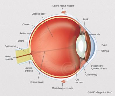
Digital painting of the anatomy of the human eye in cross-section, including the lens, pupil, iris, cornea, retina, optic nerve, sclera and vitreous body
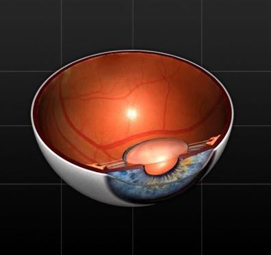
Peg Gerrity
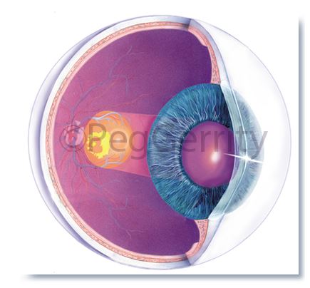
Vision loss caused by an overgrowth of choroidal blood vessels in the retina
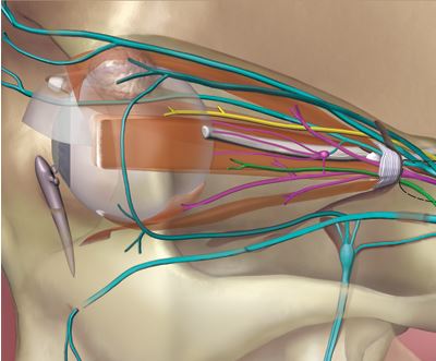
Matthew Chansky | Medical Design Agency
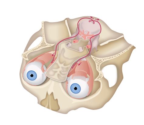
Eye and Midbrain Nerves and Muscles
Phototake – Masters in Medical Images
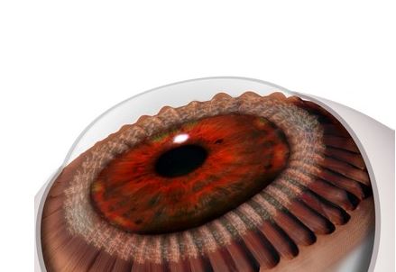
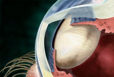
Author: Carol & Mike Werner
Eye: pars plana, part of the ciliary body
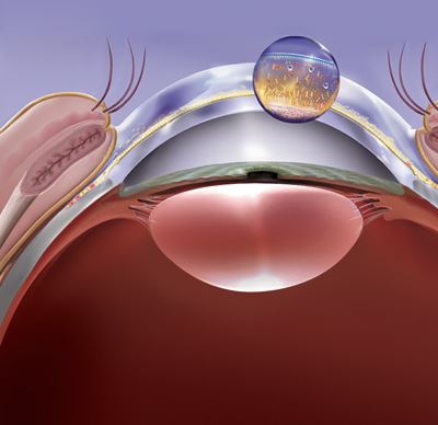
Brian Harrold | Beru Graphics
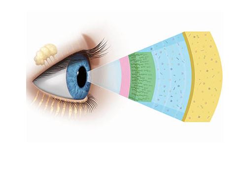
3 film layers associated with dry eye
Destry Sloane | Animated Biomedical Productions
You can explore more illustrations and animations on a wide variety of subjects, techniques, and markets using the easy and convenient Specialty Search on Medillsb.com.


