
Eye Surgery Reel – this is a collection of excerpts from Trinity’s surgical animations which depict optical or ophthamological conditions and corrective procedures:
00:00 – Close-up of the human eye.
This photo-real close-up contains a life-like representation of the frontal view of an intact and healthy human eye.
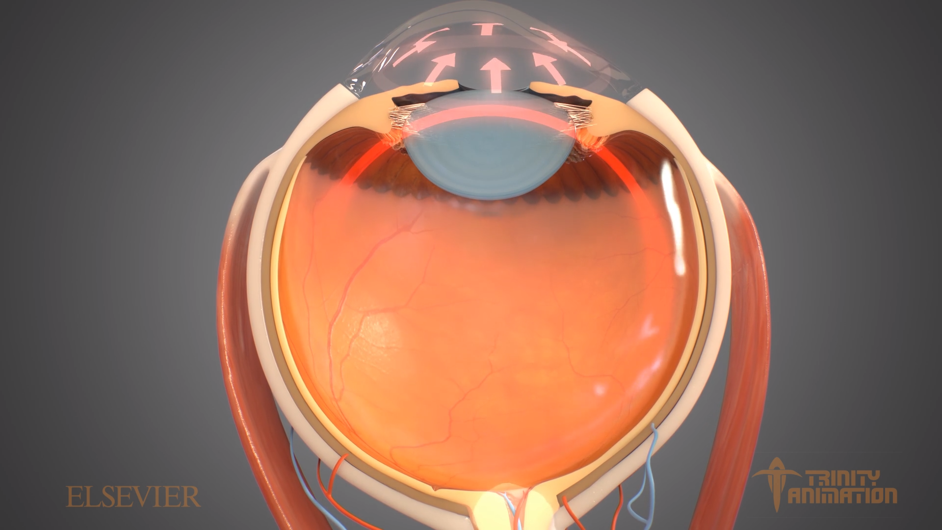
00:07 – illustration fluid build-up in open-angle glaucoma.
The source animation depicts how in open-angle glaucoma, the angle in your eye where the iris meets the cornea opens at a healthy angle, but the eye’s drainage canals become clogged over time, causing an increase in internal eye pressure and subsequent potential damage to the optic nerve.
00:17 – Lasik eye surgery procedure
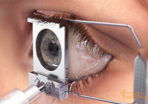
Here the surgeon uses a mechanical surgical tool called a microkeratome to create a thin, circular “flap” in the cornea. The surgeon then folds back the hinged flap to access the underlying cornea (called the stroma) and removes some corneal tissue using an excimer laser. This highly specialized laser uses a cool ultraviolet light beam to remove (“ablate”) microscopic amounts of tissue from the cornea to reshape it so it more accurately focuses light on the retina for improved vision. For nearsighted people, the goal is to flatten the cornea; with farsighted people, a steeper cornea is desired. After the laser reshapes the cornea, the flap is then laid back in place, covering the area where the corneal tissue was removed. Then the cornea is allowed to heal naturally. Laser eye surgery requires only topical anesthetic drops, and no bandages or stitches are required.
00:26 – scleral buckling application.
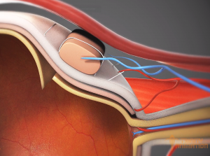
“Scleral buckling” is a surgical procedure used to repair a retinal detachment. The scleral, or the white of the eye, is the outer supporting layer of the eyeball. In this surgery, a surgeon attaches a piece of silicone or a sponge onto the white of the eye at the spot of a retinal tear. The buckle is designed to repair retinal detachment by pushing the sclera toward the retinal tear or break. The retina is a layer of tissue on the inside of the eye. It transmits visual information from the optic nerve to your brain. A detached retina shifts from its normal position. If left untreated, retinal detachment can cause permanent loss of vision.
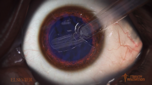
0:33 – Cataract Removal and replacement lens insertion
A cataract is an abnormality in the lens of the eye. The lens of the eye is similar to the lens of a camera—it serves to focus on objects both near and far away. To work well, the lens must be clear, With age, the lens may slowly become cloudy, so most adults who live into their 60s will eventually get at least some mild cataracts. Cataract extraction is a surgical procedure by which the cloudy natural lens of the eye is removed. The surgeon makes a small cut on the surface of the eye near the clear part of the eye called the cornea. A small instrument is inserted into the incision and the cloudy lens material is removed from the eye. The surgeon then inserts an replacement lens. This intraocular lens, also known as an implant or artificial lens, is used to help the eye focus after surgery. The lenses come in different strengths, like glasses, and the surgeon will do several measurements to decide which lens strength is right each patient
0:42 – Trochlear cranial nerve sending sensory to control eye movement
The trochlear cranial nerve is one of four cranial nerves involved in vision and movement of the eyes. Best known of these four, is the optic nerve which is the sensory nerve for vision: it transmits information from the eyes to the brain. The three remaining cranial nerves involved in vision control the eye muscles: the oculomotor nerve, which controls the majority of the muscles, the trochlear nerve, which controls the superior oblique muscle, and the abducens nerve, which controls the lateral rectus muscle. The trochlear nerve is illustrated here as it moves the eyeballs by sending nerve impulses to the superior oblique muscles which are among the group of muscles that rotate the eyeballs in their sockets. (The action of this nerve is coordinated with those of the oculomotor and abducens nerves i.e. cranial nerves III and VI.
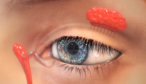
0:46 – Corneal abrasions being visualized with Fluorescein stain
Fluorescein eye stain. This is a test that uses orange dye (fluorescein) and a blue light to detect foreign bodies in the eye. This test can also detect damage to the cornea: the outer surface of the eye.
0:51 – Closeup of Eye showing Lacrimal gland generating tears
The lacrimal glands are paired, almond-shaped exocrine glands, one for each eye, that secrete the aqueous layer of the tear film. They are situated in the upper lateral region of each orbit, in the lacrimal fossa of the orbit formed by the frontal bone. Inflammation of the lacrimal glands is called dacryoadenitis.
Trinity’s surgical animations as illustrations for professional curriculum.
Surgical animations are very valuable tools for teaching difficult procedures such as eye surgeries, because of the clear and detailed visuals that they are able to generate, Trinity’s 3D renderings of eye surgeries are able to provide a view of the anatomical structures of the eye from any angle or virtual camera path. Trinity’s animations portray the surface texture of materials such as the skin, eyeball exterior, medical tools, as well as the interior parts of the eye with an unparalleled level of accuracy. Even the representation of bodily fluids match the viscosity of those found in the human body. Clearly, Trinity’s animations compare very well to other options such as live footage or medical books with 2D illustrations and diagrams,
Trinity’s surgical animations as patient education material
Clearly, live footage of these eye surgery procedures can be uncomfortable for many viewers to watch: especially patients. Trinity’s surgical animations of the procedures convey information that 2D images cannot but omit disturbing materials founds in live action recordings of the actual procedures.
Each surgical animations are created as according to the needs of Trinity’s client and is delivered royalty free for maximum usefulness and versatility.
To accurately create these anatomically correct surgical animations, clients provide Trinity animators with illustrations, diagrams, and sometimes live footage of the eye surgeries that Trinity’s animators can base their 3D models off of. Additional research is often required by Trinity animators to accurately represent the structures of the eye which they often access through medical books.
Trinity animators not only have the goal of making surgical animations anatomically correct but also keeping the viewers interested through color, movement, staging, and more. They have the ability to engage people with interesting visuals that are impactful and memorable. Occasionally, graphics are included for clarity. For instance, animated arrows are incorporated in certain shots to indicate the direction of various materials. These visual aspects of Trinity’s eye surgery animations create a clear demonstration of the procedures and resonate with the viewers in a way that doesn’t compare to any other teaching method, which is what makes eye surgery medical animations a valuable tool.
Trinity animator’s surgical animations accurately communicate the procedures to patients and medical staff members in an impactful and easily understood way. This skill allows Trinity to create professional and high-quality medical animations which communicate important information that is rather difficult to understand while holding the viewer’s attention.
- Cataract: Affects more than 24.4 million Americans by age 40.
- Glaucoma: Affects more than 2.7 million Americans by age 40.
- Corneal Transplants: 48,229 corneal trnasmplants wre performed in the U.S. in 2013. Since 1961 more than 1,000,000 men, women and children have had their sight restored through a corneal transplant.
- Color Blindness: Approximately 8% of men and 0.5% of women and among populations with Norther European ancestry have the most common form of color blindness that makes it hard to see red or green. Incidence of this is lower in almost every other population studied.
- Corrective Eyeware: More than 15 million American use corrective eyewear to compensate for refractive errors, which is due to the cornea being less than perfectly rounded.
- Contact Lenses: 37 million Americans wear contact lens.
- Lasik Surgeries: 800,000 LASIK oR PRK surgeries were performed in 2010.
The post Surgical Animations – Eye and Optical appeared first on Trinity Animation | Blog.


