
Inside the cell with Nanobot medical animation studio.
Understanding cellular architecture has always been an important issue for understanding biology. Cells contain hundreds of organelles and macromolecular assemblies which cannot be seen with optical microscope. The very first electron microscopy images of eukaryotic cells were attributed in 1945, and it took until 1949 until the cell’s internal structures were first shown when samples for the first time were embedded in plastic to enable thin sections.
Nowadays electron microscopy demonstrates fearsome advances in imaging technique such as focused ion beam scanning electron microscopy which makes it possible to generate a three-dimensional picture of the cell structure. Focused ion beam scanning electron microscopy provides a high level of detail showing many organelles in their natural state.
In 3D projects, we often show the cell organelles, and our aim is to make them realistic not only yielding to the latest visualization methods but also surpassing them.
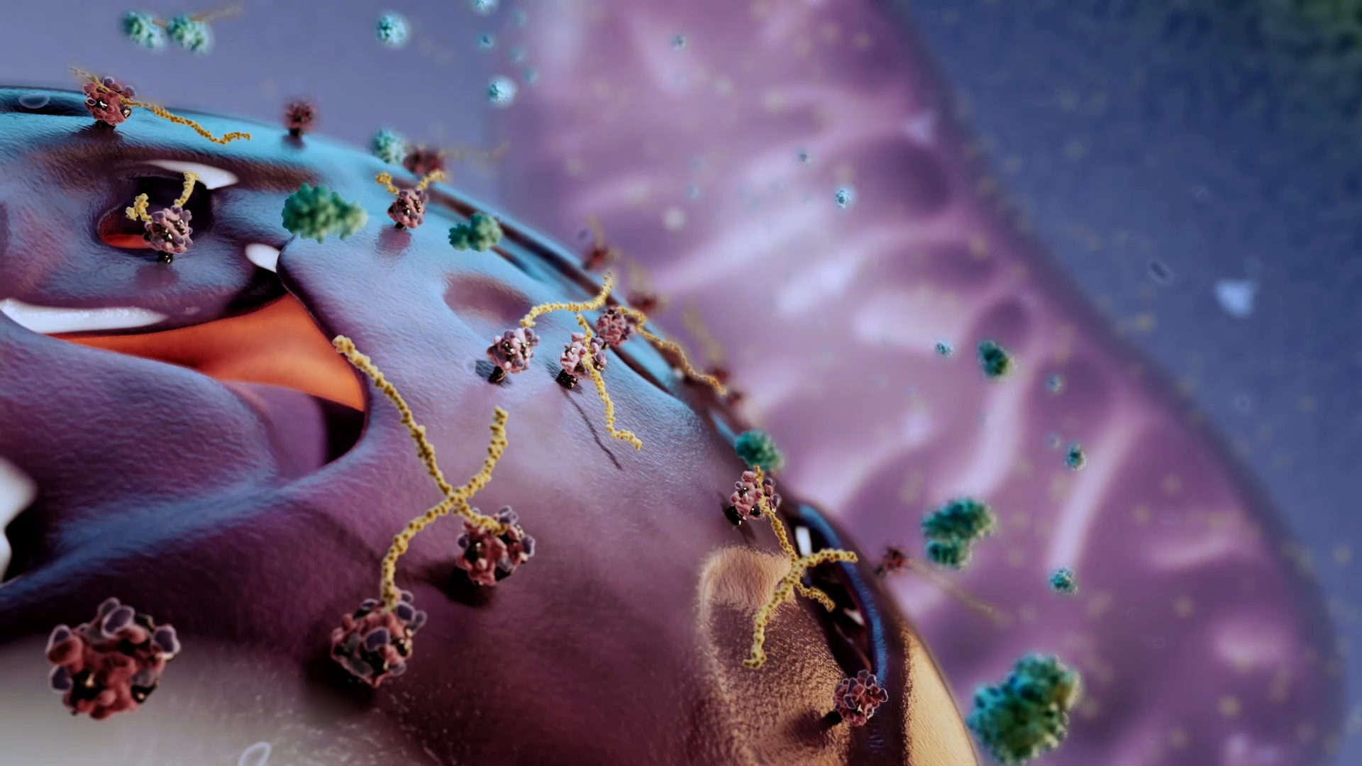
Usually, cellular organelles play role as part of pathogenesis or drug targets. Most often we model the cell membrane so it is the beginning of everything for us. The cell membrane mostly consists of a double layer of phospholipids. When we create a single phospholipid, it is important that the phospholipid doesn’t look like a tooth or a pin. That’s why we keep the modeling process under the supervision of medical advisors. Next, the animators assemble a complete membrane making it fluid and rigid. A separate kind of art is to make every model look naturally vivid. It’s not obvious, but it requires a lot of configurations.
One of the most beautiful processes associated with the cell membrane is endocytosis. Endocytosis is a cellular process in which substances are brought into the cell. Look how we show this process in 3D.
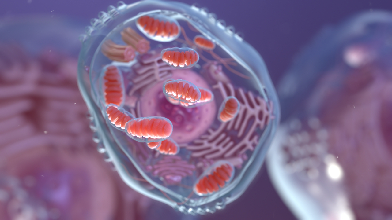
The mitochondrion is our favorite organelle. Mitochondria are found in almost all cells of the body since they produce energy molecules. Erythrocytes don’t contain mitochondria because there aren’t many energy-consuming processes. The size of mitochondria is similar to the size of many bacteria, and this is not a coincidence. According to the symbiotic theory, mitochondrion is a former bacterium that a long time ago was phagocytosed by another cell but could survive. That’s why mitochondrion has its own genome that is substantially similar to bacterial genomes. Do you want to see in AR how mitochondrion produces ATP molecules? Hurry up because ATP molecules live no more than a minute!
What do you imagine when you hear “ribosome”? Most likely, these are two ball subunits that look like a small snowman. In reality, you won’t find such a ribosome. The ribosome is mostly composed of ribosomal RNA molecules, and its shape resembles something between an oval ball and a pyramid, where you cannot distinguish the boundaries between subunits with an unaided eye. Ribosomes are about 70 times smaller than mitochondria, but every single one can synthesize any protein you can imagine.
The Golgi apparatus does not often act as a drug target but is often associated with the life cycle of many viruses, such as herpesviruses, poxviruses, and bunyaviruses. Interestingly, there is still no unambiguous theory explaining intra-Golgi trafficking of proteins. There are two major models. The first one is the vesicular transport when the Golgi cisternae remain stable. The second one is the cisternal maturation model in which the Golgi cisternae themselves function as the cargo carriers progressing from cis to trans. Anyway, both trafficking models allow the Golgi complex to play a central role in protein secretion.
Nowadays electron microscopy demonstrates fearsome advances in imaging techniques such as focused ion beam scanning electron microscopy which makes it possible to generate a three-dimensional picture of the cell structure. Focused ion beam scanning electron microscopy provides a high level of detail showing many organelles in their natural state.
Cells contain hundreds of organelles and macromolecular assemblies. Just like organs in the body, each organelle contributes in its own way to helping the cell function well as a whole.
As you can see scanning electron microscopy opens up a new side of reality, however, it cannot compete with 3D animation because 3D animation represents both reality and every whim of imagination. Quality 3D animation knows no boundaries showing any shapes, textures, and processes with multiple camera movements so that the final video resembles a high-quality blockbuster. Start your journey inside the cell with us! You won’t remain indifferent!
Download Free eBook “How much does Medical Animation explainer cost?”
- Should you choose Freelancer or Studio as a medical animation provider?
- What is the COST structure?
- Prepare BETTER for the project
- How to SAVE the budget?
- How to AVOID common mistakes?
The post Inside the cell with Nanobot Medical Animation Studio appeared first on Nanobot Medical Animation Studio.


