
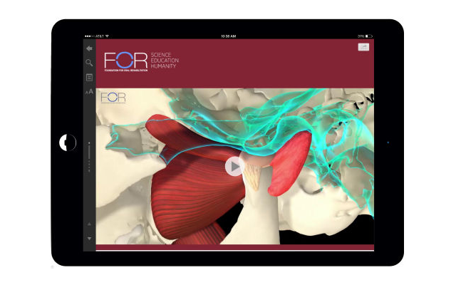
We had the pleasure to work with FOR and bring to life an exceptional resource for those desiring in-depth knowledge of temporomandibular joint anatomy.
An incredible collection of images further enhances the experience of understanding the more intricate anatomic characteristics of the TMJ.
The Temporomandibular Joint joins the existing digital textbook Single Implants and Their Restoration.
Those who study and use this digital resource will come away with an expanded appreciation of the temporomandibular joint and an enhanced ability to visualize the 3D complexity of the joint. This wonderful new book features:
- Over 650 images and illustrations, 325 scientific and literature references, 12 videos and nine animations
- A comprehensive, evidence-based review of the bony anatomy, normal and abnormal periarticular soft tissue anatomy, including extensive details about the articular disc, and muscles associated with the temporomandibular joint;
- A richly-illustrated chapter on diagnostic imaging of the TMJ showing normal and abnormal characteristics;
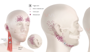
- A review of the characteristics exhibited by patients with temporomandibular disorders;
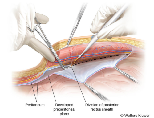
- An exceptional collection of images (bony images, cross-sectional images through cadaver specimens, histologic images, CBCT and MRI images;
- World-class animations of the joint and muscles;
- Self-assessment “Validate Your Learning” statements throughout the text and end-of-chapter self-assessment quizzes;
- A glossary that defines terms throughout the text


